Flow Imaging Microscopy
ARTiMiS
Autonomous Real-Time Microbial Scope
Complete automation for microalgae monitoring. From raw sample to actionable results, ARTiMiS delivers at a fraction of the cost of commercial alternatives.
95%
Classification Accuracy
3-5 min
Operator Time
$8.5K
Setup Cost

What ARTiMiS Does
Automated cell counting, real-time classification, and label-free phenotyping in a compact, field-deployable package.
95% Classification Accuracy
AI-powered identification with fine-tuned models for real-time tailored results.
Fully Automated
No external computer required. Load a sample, configure parameters, retrieve results.
Validated Performance
700+ sample, 24-month industrial study. More accurate than probes and gold standard FIM instruments.
Label-Free Analysis
Characterize phenotypes and assess cell health without fluorescent dyes.
How It Compares
ARTiMiS delivers industry-leading results at a fraction of the cost.
| Capability | Manual Microscopy | Commercial FIM/IFC | ARTiMiS |
|---|---|---|---|
| Setup Cost | $2K–$10K | $50K–$150K+ | $8.5K |
| Sample Time (Full Lifecycle) | 60–90 min | 15–60 min | <30 min |
| Operator Time/Sample | 60–90 min | 30+ min (curation) | 3–5 min |
| Classification | Manual (expert required) | Semi-automated | Fully automated |
| External Computer | Not needed | Required | Not needed |
| Real-time Results | No | Partial | Yes |
| Deployment | Lab only | Lab only | Lab, at-line, or field |
Savings Calculator
See how quickly ARTiMiS pays for itself based on your current workload.
Hours Saved Weekly
9.2
Value Saved Monthly
$1,190
Value Saved Annually
$14,290
Breakeven
7.1 months
Based on $8,499 ARTiMiS system cost and 90% average reduction in operator time.
Validated Performance
Rigorously validated through peer-reviewed research and real-world deployment.
Head-to-Head vs. FlowCam
In a 24-month industrial study with daily sampling (700+ samples):
Biomass Accuracy (R², higher is better)
0.74 vs 0.41
RMSE (lower is better)
0.68 vs 1.00
Classification Accuracy

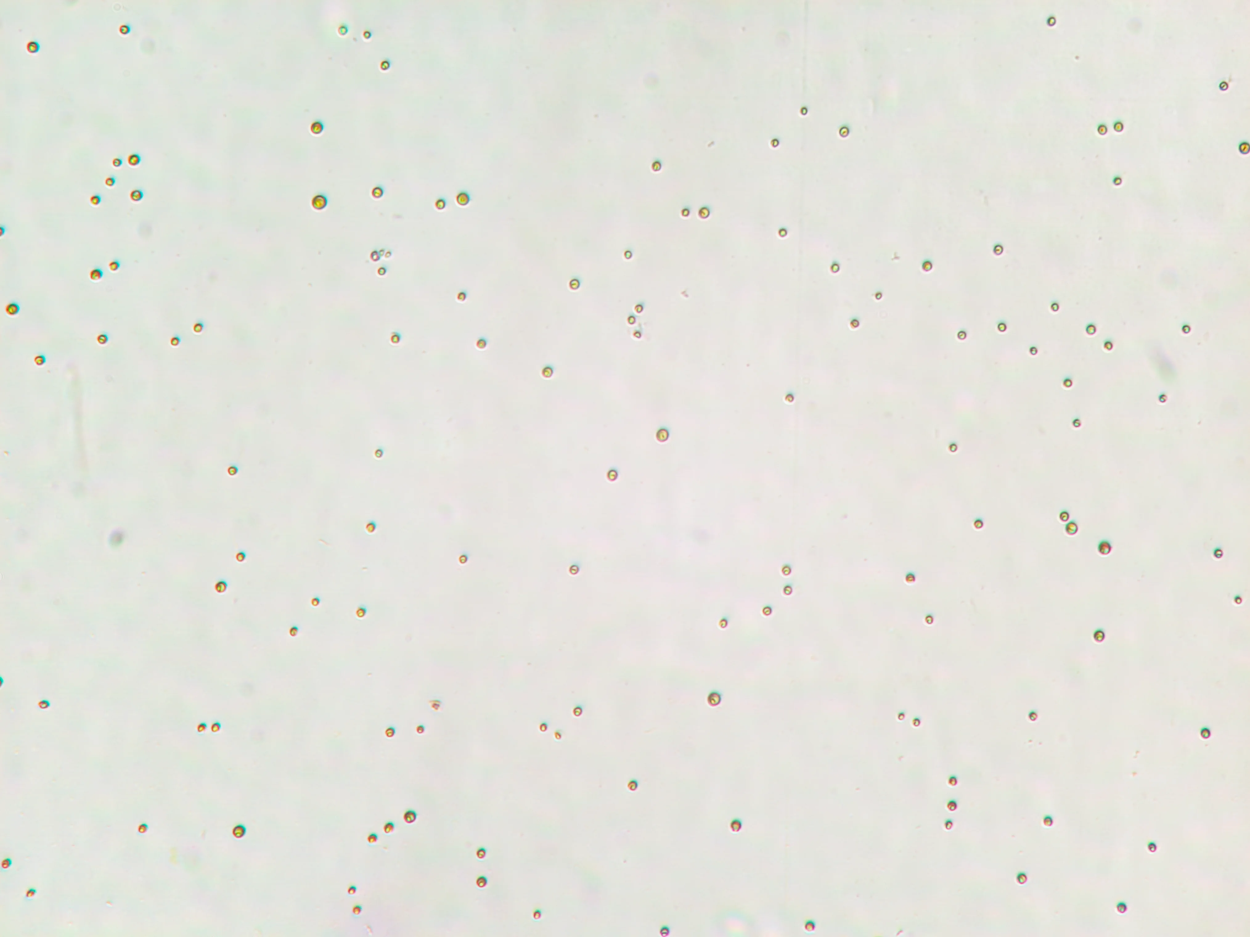
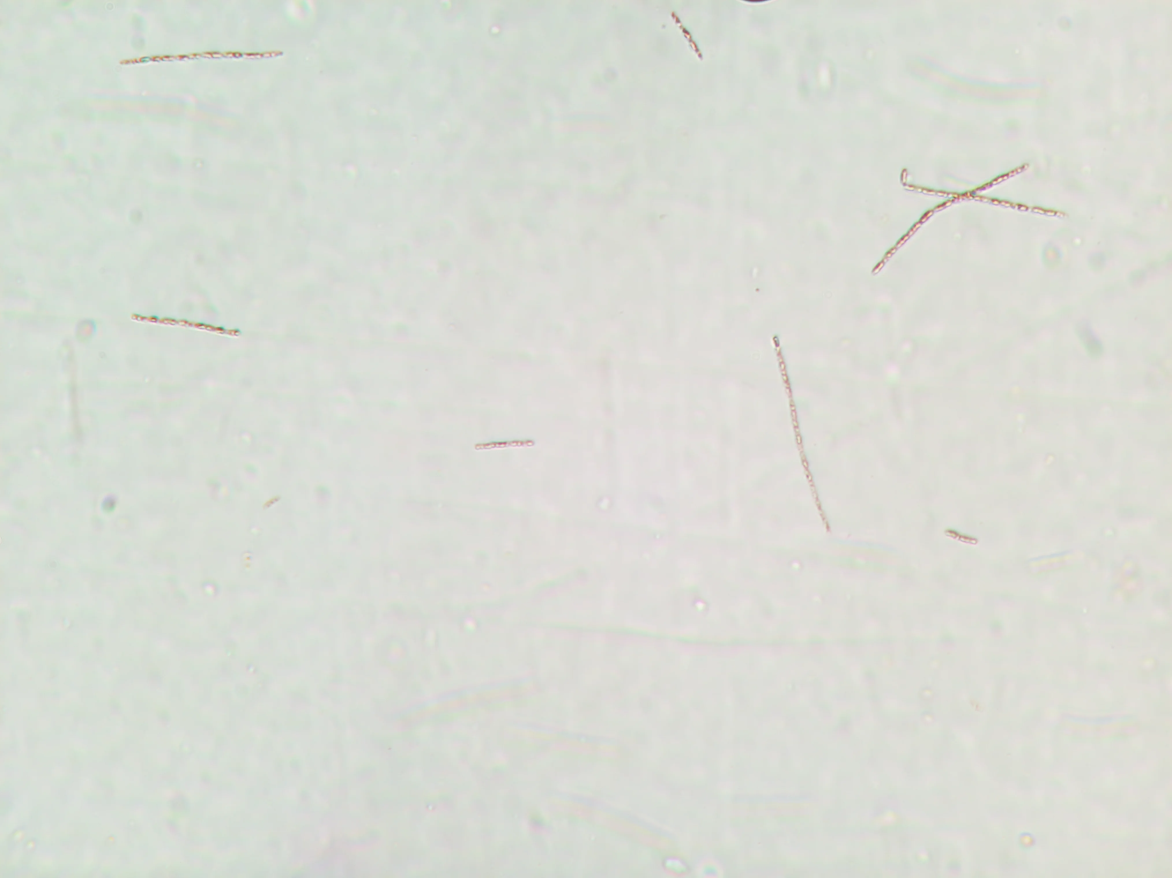
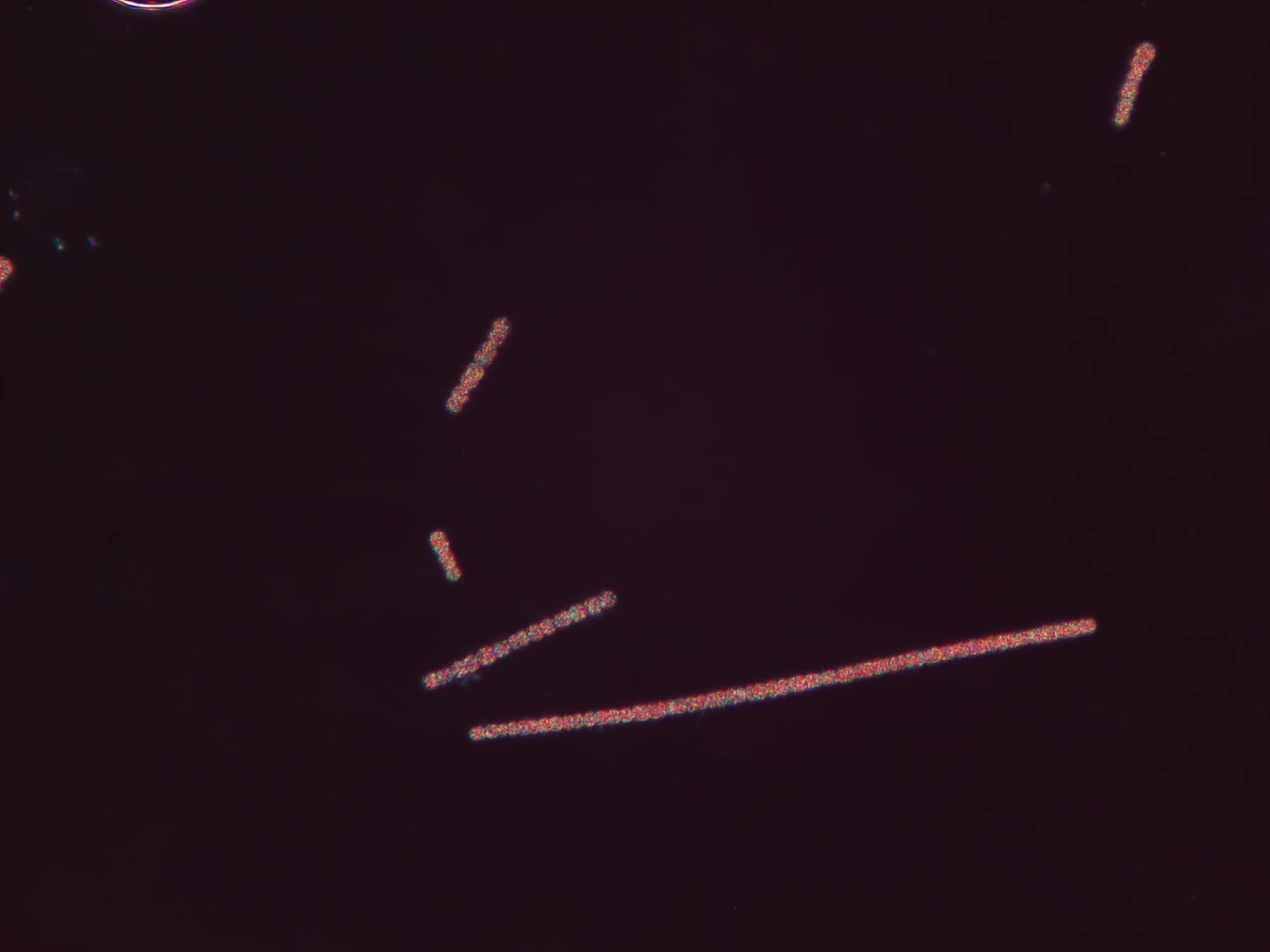
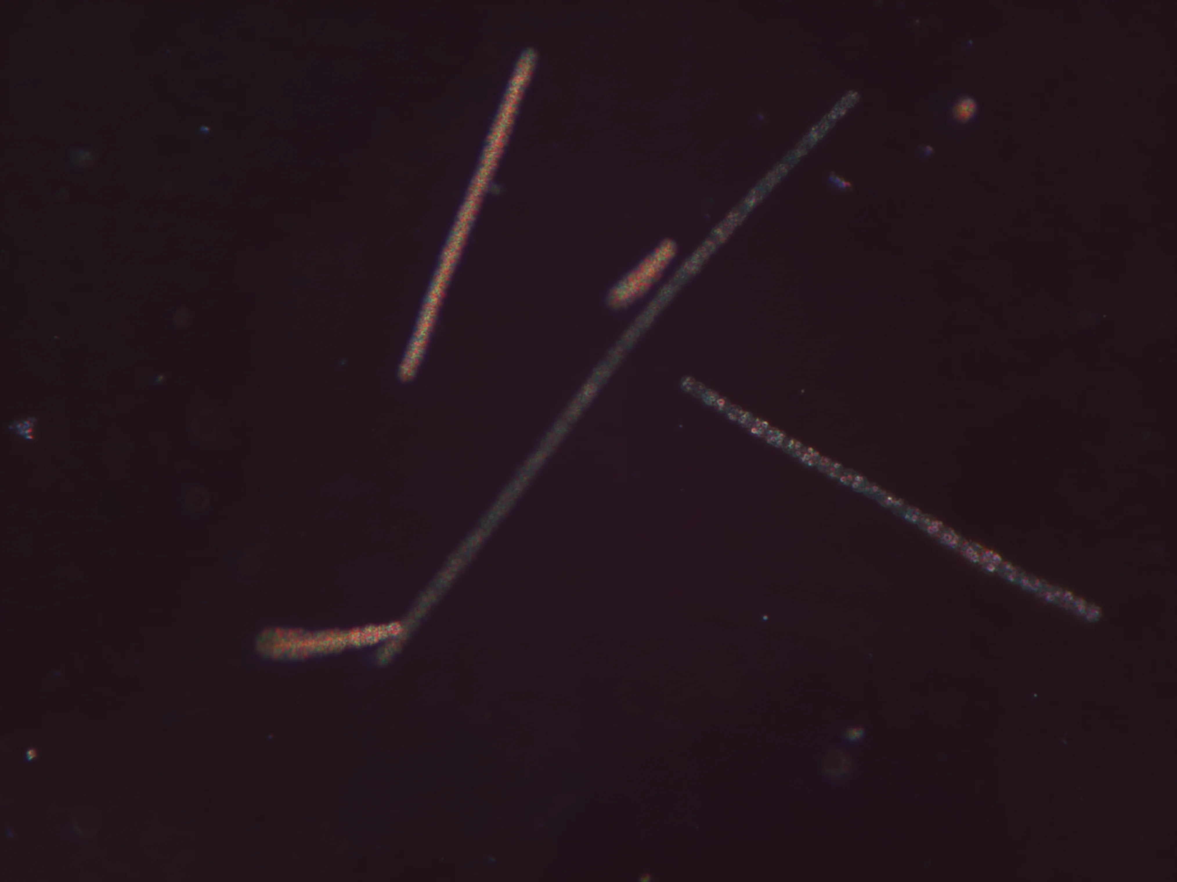
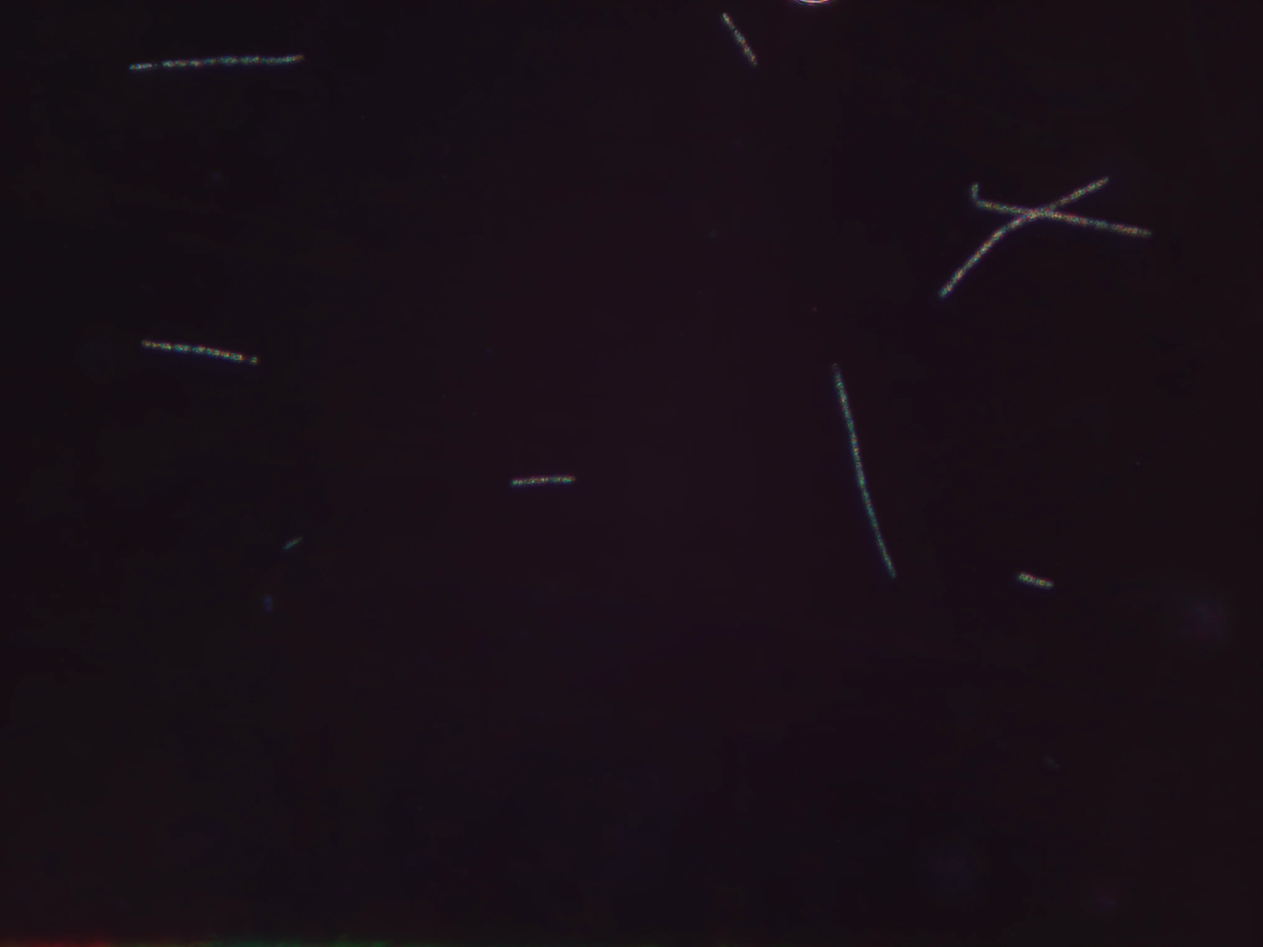
High-Resolution Widefield Imaging
ARTiMiS captures images with a wide field-of-view at a 12MP resolution, and can do so in both brightfield and darkfield illumination formats.
Brightfield Illumination
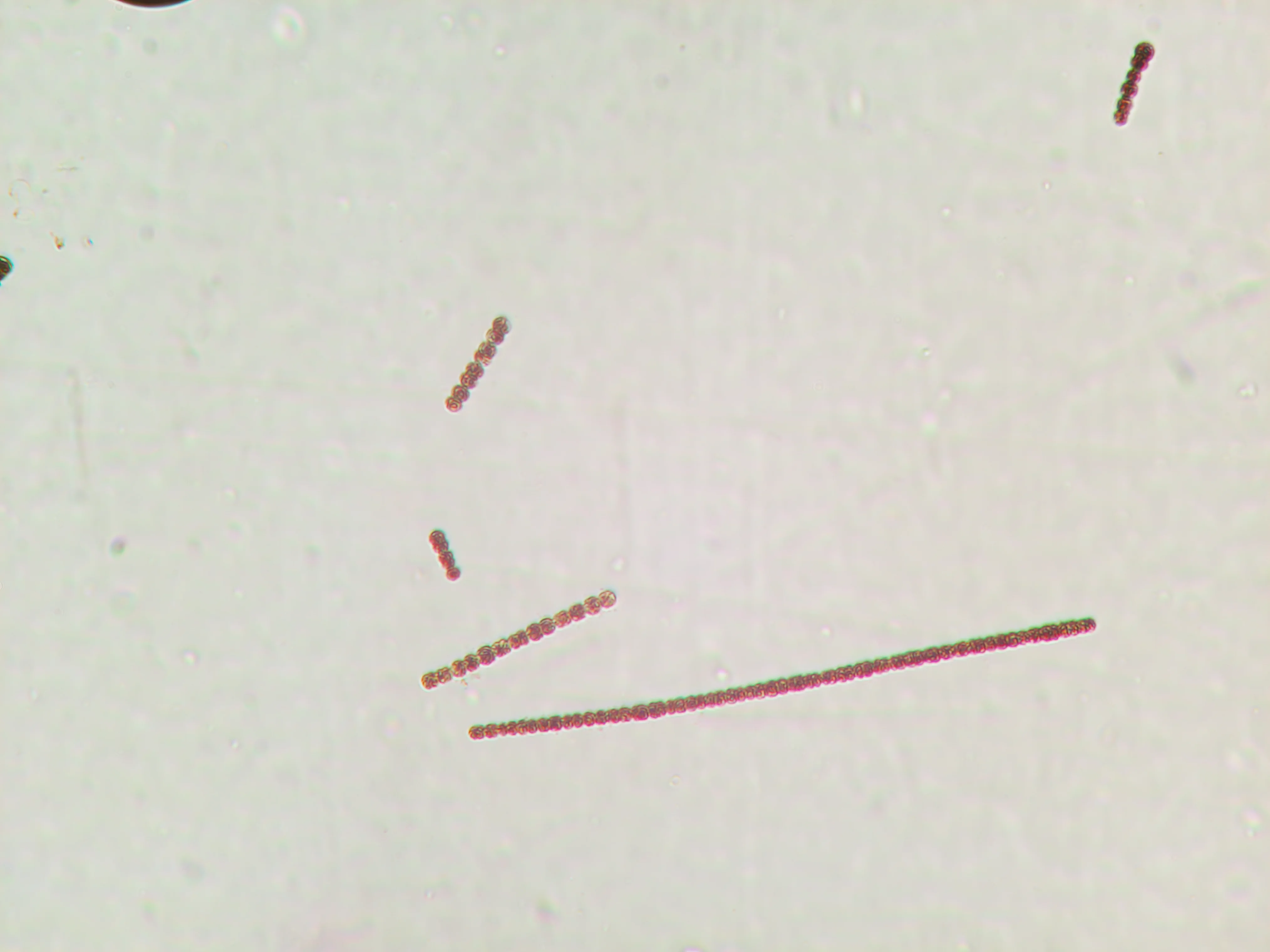
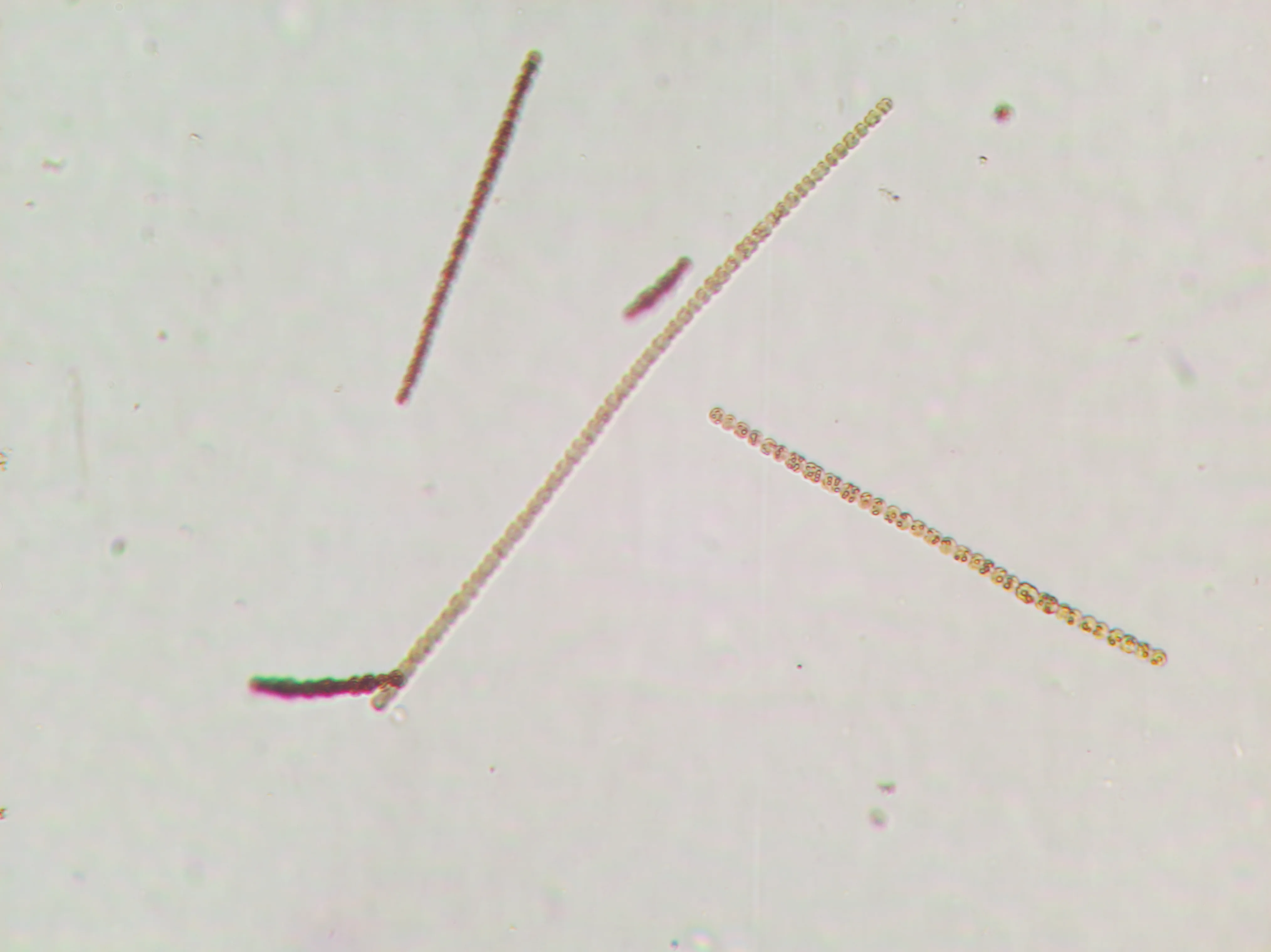
Dolichospermum
Darkfield Illumination
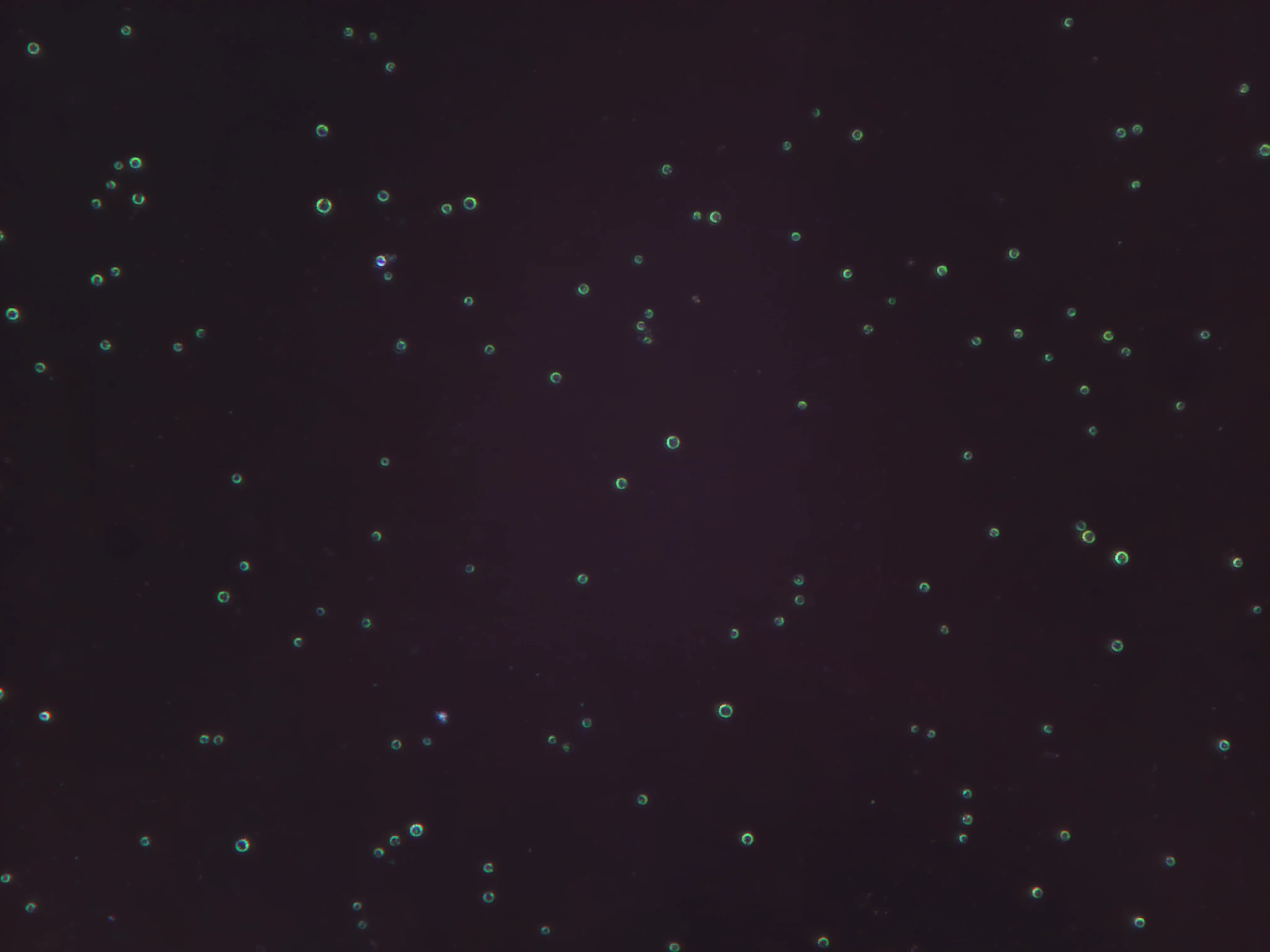
Dolichospermum
Applications
From industrial cultivation to environmental monitoring, ARTiMiS adapts to your workflow.
Industrial Microalgae Cultivation
- Monitor culture purity and detect contamination early
- Optimize harvest timing based on growth phase
- Track phenotype changes without fluorescent labels
- Prevent culture crashes that cost $10K–$1M+ per event
Wastewater Treatment
- Real-time biomass composition monitoring
- Identify key taxa driving nutrient removal performance
- Optimize process control parameters
- Meet tightening phosphorus discharge limits
Drinking Water Management
- Early warning for harmful algal blooms (HABs)
- Taxonomic identification for treatment optimization
- Source water quality tracking
- Reduce public health risk
Research & Environmental Monitoring
- High temporal resolution ecological studies
- Climate change impact assessment
- Long-term phytoplankton community tracking
- Eliminate personnel bottlenecks
Technical Specifications
Complete specifications for the ARTiMiS flow imaging microscope system.
Physical
- Dimensions
- 150 × 230 × 115 mm (6 × 9 × 4.5 in)
- Weight
- 949 g (2.1 lb)
- Power Input
- 12V DC, 5A (60W max)
Optical System
- Illumination Modes
- Brightfield, Darkfield
- Effective Magnification
- 8×
- Spatial Resolution
- 0.2 μm/pixel (5 pixels per micron)
- Minimum Particle Size
- 2.0 μm
- Camera Sensor
- 12.3 MP
- Image Resolution
- 4056 × 3040 pixels
- Field of View
- ~810 × 610 μm
- Imaging Volume
- 0.1 μL per frame
Onboard Computing & Connectivity
- Processor
- Broadcom BCM2712, 2.4 GHz Quad-Core ARM Cortex-A76
- Memory
- 8 GB LPDDR4X SDRAM
- Storage
- 500 GB SSD
- Operating System
- Linux (Raspberry Pi OS)
- Wi-Fi
- Dual-band 802.11ac (2.4 GHz / 5 GHz)
- Bluetooth
- 5.0 (BLE)
- Ethernet
- Gigabit
- USB
- 2× USB 3.0, 2× USB 2.0
Sample Handling
- Minimum Sample Volume
- 1.0 mL (recirculation mode)
- Flow Configuration
- Stop-flow with particle settling
- Settling Time
- Configurable
- Microfluidic Channel
- 200 μm depth, TOPAS (COC) polymer
- Optimal Concentration
- 10⁵–10⁶ cells/mL
- Validated Range
- 10⁴–10⁷ cells/mL
Detection & Quantification Limits
- Limit of Detection (LoD)
- 3.2×10³ particles/mL
- Limit of Quantification (LoQ)
- 5×10³ particles/mL
- Dynamic Range
- 10³–10⁷ cells/mL
- Minimum Detectable Particle
- 0.6 μm diameter
- Minimum Measurable Particle
- 2.0 μm diameter
Environmental
- Housing Material
- 3D-printed ASA, PLA, PETG
- Operating Temperature
- 4–40°C
- Operating Humidity
- 20–80% RH (non-condensing)
- IP Rating
- IP52
Data Export Protocols
| Protocol | Format | Export Medium |
|---|---|---|
| Direct File Export | CSV, JSON, PNG, JPG, TIFF | USB drive, cloud upload |
| WebSocket | JSON | Real-time data streaming |
| Modbus TCP/UDP | Standard registers | SCADA/PLC integration |
Exported data includes: raw particle images, 50+ morphological parameters per particle, classification predictions with confidence scores, and aggregated sample statistics.
Maintenance & Consumables
- Channel Rinse
- Every sample (recommended)
- Preventative Cleaning
- Daily (recommended)
- Tubing Replacement
- ~300 samples or 3 months
- Microfluidic Channel Swap
- ~300 samples
- Microfluidic Chip Replacement
- ~3000 samples
- Software Updates
- Automatic (Wi-Fi)
Ready to Automate Your Microalgae Monitoring?
See ARTiMiS in action: schedule a demo and send us a sample!
Questions? Contact us at info@skopii.com